
nCounter®
Neuropathology Panel
Helping Your Research
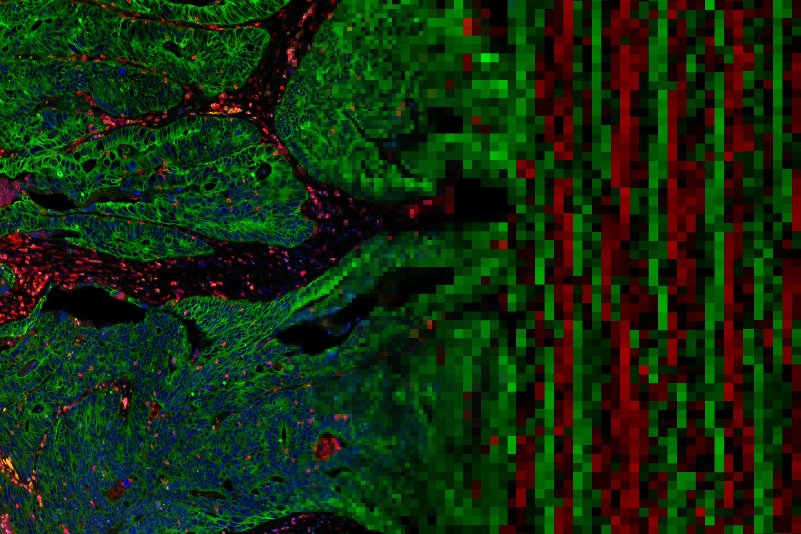
Biomarker discovery and signature development are essential for identification of novel therapies and for earlier detection and development of therapies for neurodegenerative disorders like Alzheimer’s Disease (AD), Parkinson’s Disease (PD), and Amyotrophic Lateral Sclerosis (ALS). The nCounter Neuropathology Panel helps perform comprehensive multiplex gene expression analysis in human or mouse samples with genes involved in six fundamental themes of neurodegeneration: neurotransmission, neuron-glia interaction, neuroplasticity, cell structure integrity, neuroinflammation, and metabolism.
- Developed for research of Alzheimer’s Disease, Parkinson’s Disease, Amyotrophic Lateral Sclerosis, Frontotemporal Dementia, Huntington’s Disease, and other neurological disorders
- Includes unique cell typing feature for measuring the abundance of five important CNS cell types, including neurons, astrocytes, microglia, oligodendrocytes, and endothelial cells


Panel Selection Tool
Find the gene expression panel for your research with Panel Pro
Find Your PanelProduct Information
Genes included in the Neuropathology Panels provide unique cell profiling data for measuring the abundance1 of five important cell types including neurons, astrocytes, microglia, oligodendrocytes, and endothelial cells. The table below summarizes each cell type represented in the panels along with the gene content qualified through current literature references.
1Danaher P. et al. Gene expression markers of Tumor Infiltrating Leukocytes JITC 2017
Functional annotations for 23 fundamental pathways and processes were assigned across all genes in the Neuropathology Panels allowing for a practical view of important aspects of the onset and progression of neurodegenerative disease.
Related Resources


Publications
Spatial transcriptomics reveals molecular dysfunction associated with cortical Lewy pathology
A key hallmark of Parkinson’s disease (PD) is Lewy pathology. Composed of α-synuclein, Lewy pathology is found both in dopaminergic neurons that modulate motor function, and cortical regions that control cognitive function.
Digital spatial profiling of segmental outflow regions in trabecular meshwork reveals a role for ADAM15
In this study we used a spatial transcriptomics approach to identify genes specifically associated with either high or low outflow regions in the trabecular meshwork (TM) that could potentially affect aqueous humor outflow in vivo. High and low outflow regions were identified and isolated from organ cultured human anterior segments perfused with fluorescently-labeled 200 nm FluoSpheres.
Clinically relevant molecular hallmarks of PFA ependymomas display intratumoral heterogeneity and correlate with tumor morphology
Posterior fossa type A (PF-EPN-A, PFA) ependymoma are aggressive tumors that mainly affect children and have a poor prognosis. Histopathology shows significant intratumoral heterogeneity, ranging from loose tissue to often sharply demarcated, extremely cell-dense tumor areas.
Request a Quote
Contact our helpful experts and we’ll be in touch soon.