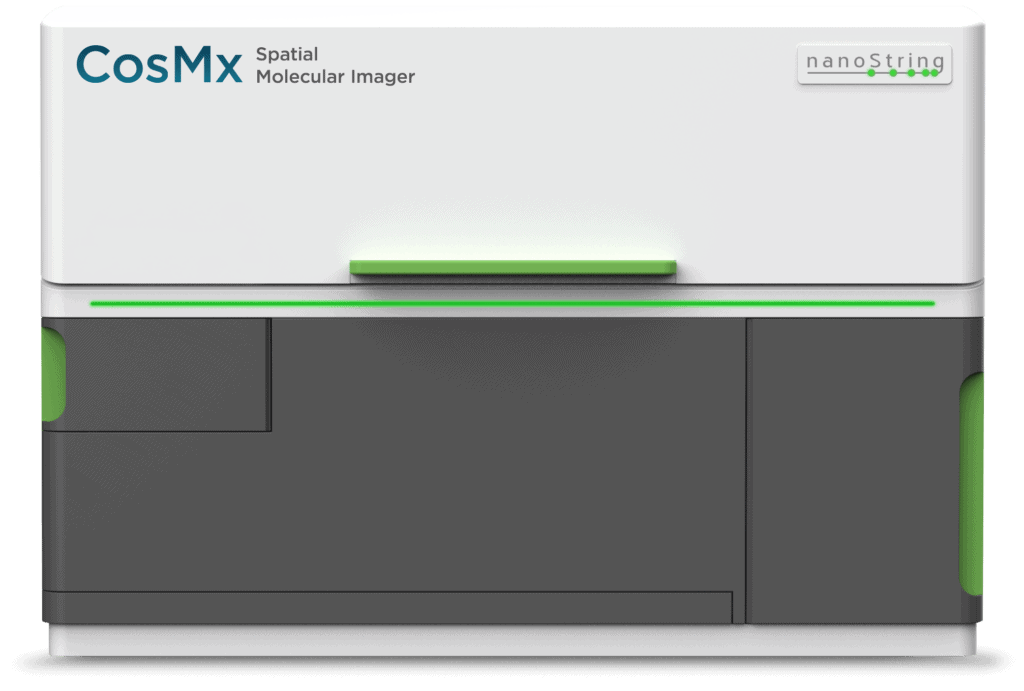
Pioneering Spatial Transcriptomics: Exploring Single-Cell Imaging with the CosMx SMI
In Collaboration With:

The CosMx SMI is accessible to researchers at CWRU through the Light Microscopy Imaging Core (SOMLMIC) for project execution.
Researchers at Case Western Reserve University are invited to join us for an upcoming seminar highlighting the capabilities and broad applications of the CosMx SMI, the first high-plex in situ analysis platform to provide spatial multiomics with formalin-fixed paraffin-embedded (FFPE) and fresh frozen (FF) tissue samples at cellular and subcellular resolution.
The session will also highlight the newest breakthrough in SMI technology, the CosMx™ Human 6K Discovery Panel enabling scientist to profile up to 6,000 RNA transcripts. Dr. Jenkins, director of the School of Medicine Light Microscopy and Imaging Core Facility will share the services available through the core facility, and the process for CWRU researchers to jumpstart their CosMx SMI projects.
Examples of Applications
The CosMx Spatial Molecular Imager (SMI) offers a versatile solution trusted by esteemed researchers for single-cell imaging across diverse applications. Here are several examples highlighting its relevance across various research domains:

- Oncology: Identify cellular neighborhoods that reveal tumor heterogeneity at single cell resolution
- Neuroscience: Understand how CNS cells interactive with one another in health and disease
- Infectious Disease: Profile the immune response at the single cell and subcellular level
- Immunology: Assess heterogeneity of immune cell population and disease progression at the cellular and sub-cellular level.
- Developmental Biology: Reveal highly heterogeneous but small cell populations and relationships from the earliest stages of development.
Speakers

Seth Meyers
District Sales Manager, NanoString Technologies


