GeoMx® Immune Pathways Panel
Helping Your Research
Targeted Content for Cancer Biology
The GeoMx Immune Pathways Panel is designed for targeted profiling of the tumor, tumor microenvironment, and tumor immune status.
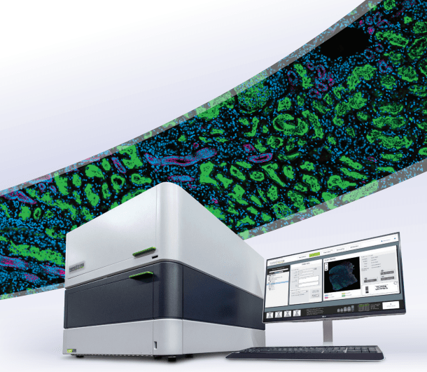
Profile up to 96 RNA targets with spatial resolution from a single tissue section using the GeoMx Digital Spatial Profiler (DSP).


How it Works
The Immune Pathways panel contains 84 targets plus controls designed for broad coverage of the tumor and tumor microenvironment. GeoMx RNA assays contain in situ hybridization (ISH) probes conjugated to unique DNA indexing-oligonucleotides via a UV-photocleavable linker. After region of interest (ROI) selection on GeoMx DSP and UV cleavage of the oligonucleotides, each DNA oligonucleotide is recognized by a unique Reporter probe that contains a fluorescent barcode. Reporter probes are imaged and counted by the nCounter® Analysis System to provide a direct, digital readout of spatially resolved RNA expression.
- Curated content designed for immuno-oncology research
- Includes tumor and tumor microenvironment coverage plus the Tumor Inflammation Signature (TIS)
- Pre-validated in multiplex format for use in human FFPE or fresh frozen tissue
- Compatible with RNAscope® and antibody morphology markers for tissue imaging
- Customizable with up to 10 additional targets of interest
- For use with nCounter readout and compatible with DSP Data Center
Curated Content for Immuno-Oncology
The GeoMx Immune Pathways panel is designed to profile key aspects of the tumor and tumor microenvironment biology.
- Profile the global immune response
- Assess microenvironment immune activity
- Quantify tumor reactivity
- Measure the 18-gene Tumor Inflammation Signature known to be associated with response to PD-1/PD-L1 inhibitor pathway blockade
Accompanying Morphology Marker Kits are available for tissue visualization and ROI selection.


Publications
Assessing Longitudinal Treatment Efficacies and Alterations in Molecular Markers Associated with Glutamatergic Signaling and Immune Checkpoint Inhibitors in a Spontaneous Melanoma Mouse Model
Previous work done by our laboratory described the use of an immunocompetent spontaneous melanoma-prone mouse model, TGS (TG-3/SKH-1), to evaluate treatment outcomes using inhibitors of glutamatergic signaling and immune checkpoint for 18 weeks. We showed a significant therapeutic efficacy with a notable sex-biased response in male mice.
Spatial transcriptomics reveals molecular dysfunction associated with cortical Lewy pathology
A key hallmark of Parkinson’s disease (PD) is Lewy pathology. Composed of α-synuclein, Lewy pathology is found both in dopaminergic neurons that modulate motor function, and cortical regions that control cognitive function.
Digital spatial profiling identifies molecular changes involved in development of colitis-associated colorectal cancer
Objective: Chronic colonic inflammation seen in inflammatory bowel disease (IBD) is a risk factor for colorectal cancer (CRC). Colitis-associated cancers (CAC) are molecularly different from sporadic CRC.
Request a Quote
Contact our helpful experts and we’ll be in touch soon.


