nCounter®
PanCancer Progression Panel
Helping Your Research
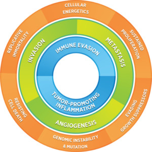
Cancer progression involves multiple processes and mechanisms that are highly interconnected. The nCounter PanCancer Progression Panel lets you perform multiplex gene expression analysis with 770 genes from each step in the cancer progression process including: angiogenesis, extracellular matrix remodeling (ECM), epithelial-to-mesenchymal transition (EMT) and metastasis.
- Comprehensive gene expression analysis of cancer progression
- Quantify gene expression of metastatic growth and suppressor genes
- Rapidly and easily screen samples for biomarker discovery or drug mechanism of action to support your research
- Customizable with up to 55 additional user-defined genes with Panel Plus option
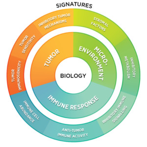
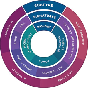
Inspired by systems biology approaches to cancer research, NanoString’s 360 Series Panel Collection gives you a 360° view of gene expression by combining carefully-curated content involved in the biology of the tumor, microenvironment, and the immune response into a single holistic assay. Each panel contains the 18-gene Tumor Inflammation Signature (TIS) that measures a peripherally-suppressed, adaptive immune response and has been shown to correlate with response to checkpoint inhibitors.
360 Series Panel Collection
Gene Coverage
Processes, features and key genes included in the panel:
-
PRIMARY TUMOR
↓ O2, ↑ Glycolysis -
DISTANT TUMOR
-
Extracellular Matrix Remodeling
The extracellular matrix (ECM) is an assembly of proteins and sugars that surrounds cells in solid tissues, primarily providing mechanical structure to cells. The ECM must be remodeled in order to accommodate tumor growth. Tumor cells rely on the MMP and LOX gene families to change the surrounding EMC environment. Several of these remodeling genes have further roles in progression, acting as transcription factors in metastatic growth.
Genes: LOXs MMPs TIMPs ITGAs ITGBs COLs SERPINs -
Epithelial-to-Mesenchymal Transition
The epithelial-mesenchymal transition (EMT) is a process that cancer cells undergo which promotes metastatic progression. Epithelial cells are defined by their ability to laterally tether to each other in sheets using intercellular junctions, where as mesenchymal cells are more elongated for motility and rely on focal adhesions for attachment. The epithelial-mesenchymal transition (EMT) is a dynamic spectrum between these two states that can be reversible depending on the surrounding processes
Genes: ZEBs SNAIs TWISTs PRRZ1 SMADs BMI1 ESR1 GSC MITF SIP1 -
Metastasis
Metastasis is a collection of cellular processes commencing when a cell migrates from the primary tumor to successful development of a distant tumor. While all of the general processes that are not included in the major three themes have been grouped in to the term ‘metastasis’. This term includes general cell growth signaling pathways, hypoxia response, and metabolic changes. Additionally, there are a set of metastasis suppressor genes that each potential new tumor site must avoid.
Genes: HIF1A BRMS1 TXNIP MED23 DLC1 CDH1 GSN KISS1 KDM1A NME1 MTOR -
Angiogenesis
Angiogenesis is the biological process of generating new blood vessels from pre-existing vasculature. In the disease state, angiogenesis is induced quite early in cancer development, a process often referred to as turning on an “angiogenic switch.” Angiogenesis is of particular interest in cancer progression considering that distant tumors cannot grow more that 2-3 cubic mm without vasculature.
Genes: VEGFs FGFs FGFRs PDGFs ANG ANGPTs EGF HGF

Panel Selection Tool
Find the gene expression panel for your research with Panel Pro
Find Your PanelProduct Information
360 Series Product Comparison
Fully-annotated gene lists in Excel format are available for each of the 360 Panels. The table below compares the biology coverage of the 360 Panels across the tumor, microenvironment, and the immune response to that of the PanCancer Panels Collection.

Publications
Assessing Longitudinal Treatment Efficacies and Alterations in Molecular Markers Associated with Glutamatergic Signaling and Immune Checkpoint Inhibitors in a Spontaneous Melanoma Mouse Model
Previous work done by our laboratory described the use of an immunocompetent spontaneous melanoma-prone mouse model, TGS (TG-3/SKH-1), to evaluate treatment outcomes using inhibitors of glutamatergic signaling and immune checkpoint for 18 weeks. We showed a significant therapeutic efficacy with a notable sex-biased response in male mice.
Spatial transcriptomics reveals discrete tumour microenvironments and autocrine loops within ovarian cancer subclones
High-grade serous ovarian carcinoma (HGSOC) is genetically unstable and characterised by the presence of subclones with distinct genotypes. Intratumoural heterogeneity is linked to recurrence, chemotherapy resistance, and poor prognosis.
Spatially Segregated Macrophage Populations Predict Distinct Outcomes In Colon Cancer
Tumor-associated macrophages are transcriptionally heterogeneous, but the spatial distribution and cell interactions that shape macrophage tissue roles remain poorly characterized. Here, we spatially resolve five distinct human macrophage populations in normal and malignant human breast and colon tissue and reveal their cellular associations.
Request a Quote
Contact our helpful experts and we’ll be in touch soon.


