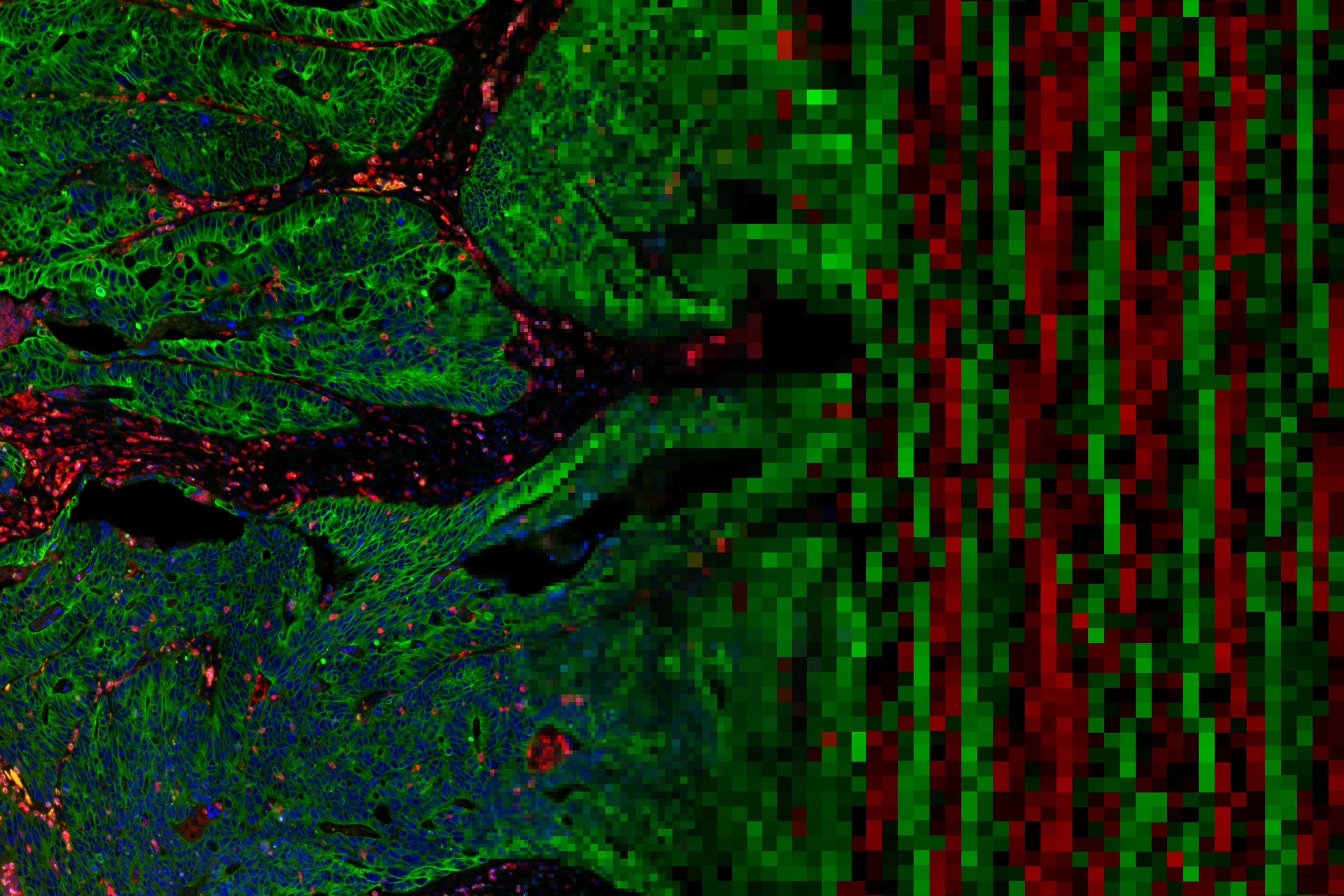
SITC 2019 may be over, but the era of Spatial Genomics enabled by the GeoMx Digital Spatial Profiler has just begun.
The curtains have fallen on SITC 2019—the 34th annual meeting organized by the Society for Immunotherapy of Cancer—but excitement amongst the participants remains high after five days of knowledge exchange and networking with the brightest stars in the field of cancer immunotherapy.
Rising young investigators and career researchers shared their latest discoveries, and NanoString’s cutting-edge GeoMx™ Digital Spatial Profiler (DSP) was showcased in multiple oral and poster sessions that highlighted the use of the platform for multiplex characterization of RNA and protein within two dimensions on a tissue section, unlocking crucial biology hidden within the spatial context of a tumor biopsy that is otherwise lost due to so-called ‘grind and bind’ bulk analysis.
At the start of the meeting on Thursday, Nov 7th, Sergio Rutella, MD, Ph.D., FRCPath from Nottingham Trent University, took the podium to present his work on the correlation between tumor infiltration and TP53 mutational status in a multi-cohort acute myeloid leukemia (AML) study.
Dr. Rutella’s group investigated the correlation of prognostic molecular lesions, focusing on TP53 mutations, with the AML immune milieu to identify a patient subgroup that would benefit from immunotherapy. Cohorts from the Cancer Genome Atlas (TCGA) and Beat AML Master Trial were analyzed in silico for immune cell type-specific and biological activity scores. TP53-mutated cases demonstrated higher levels of T cell infiltration, immune checkpoints, and interferon-γ (IFN-γ) signaling in the tumor microenvironment (TME). In contrast to patients with somatic TP53 mutations clustered in the immune-infiltrated subgroup, most TCGA-AML cases with favorable/intermediate-risk molecular lesions were classified as immune-depleted. In a subsequent study, the GeoMx DSP, in conjunction with the nCounter® PanCancer IO 360™ Gene Expression Panel was used to analyze primary AML blasts from newly diagnosed patients. These samples were found to be enriched for IL-17, tumor necrosis factor (TNF) and IFN signaling molecules as well as for T cells and immune checkpoints. This study provides evidence for a correlation between IFN-γ-dominant AML subtypes and TP53 mutations as an adverse prognosticator.
If a tumor lives within its own microenvironment, why not study it there?
Cancer cells live and prosper within a microenvironment and constantly interact with it. It is only logical to study these interactions where they happen. Anushka Dikshit, Ph.D., Jon Zugazagoitia, MD, and their respective colleagues did just that in two studies that used GeoMx DSP for biomarker identification.
In her poster, Dr. Dikshit showed how she used gene expression profiling to identify predictive biomarkers for stratification through the characterization of immune cells within the TME at the molecular level. In order to achieve this, she and her colleagues developed a robust workflow that combines the single molecule, cell, and visualization capabilities of the RNAscope in situ hybridization (ISH) assay with the highly multiplexed spatial profiling capabilities of the GeoMx DSP RNA assay. By applying both technologies to the same tissue section, her team compared differentially expressed genes within T and B cell-enriched regions of interest (ROI) and showed that while the expression of the immunoregulatory molecules CTLA4, PD-L1, PD-1, and ICOSLG were detected in both types of ROIs, the T cell-enriched ROI demonstrated significantly higher expression of these checkpoint markers. Compared to the B cell-enriched ROI, the T cell-enriched ROI also exhibited an increased inflammatory signature evidenced by elevated levels of cytokines and chemokines such as CCL5, CXCL9, and IFNG. Dr. Dikshit’s poster demonstrated that RNAscope ISH technology combined with GeoMx DSP enables users to precisely profile tissue biology at high-plex while retaining the morphological context of heterogeneous tumors.
Dr. Zugazagoitia and team ran a discovery study that demonstrated the potential of the GeoMx™ DSP for identifying spatially-resolved biomarkers associated with a favorable outcome for Non-Small Cell Lung Cancer (NSCLC) tumors treated with PD-1 checkpoint blockade therapy. In this study, the GeoMx Human IO Protein Panel (comprised of 39 markers from the GeoMx Immune Cell Profiling Core, an IO Drug Target Module, an Immune Activation Status Module, and morphology markers) was used to spatially profile protein expression. By combining the benefits of high-plex profiling with the resolution of spatial information, six immune markers associated with longer PFS (VISTA, CD127, CD56, CD4, ARG1, CTLA4) and eight markers with OS benefit (STING, CD56, CD68, CD4, B2M, CD3, ICOS, CD45) were identified from 156 protein variables per case. Expression of CD56 and CD4 in the CD45 compartment was associated with all favorable clinical outcomes in immunotherapy-treated NSCLC. GeoMx DSP data was orthogonally validated by multiplex immunofluorescence staining and cell count quantification.
Location, Location, Location
Finally, our very own Sarah Church, Ph.D. presented the results of a study on spatial profiling microsatellite instability/stability (MSI/MSS) and mismatch repair deficiency (dMMR) in colorectal cancer (CRC). In this elegant study, whole tissue gene expression analysis was conducted using the PanCancer IO 360 Gene Expression Panel and the MSI/dMMR status and Tumor Inflammation Signature (TIS) score were quantified for 48 CRC tumors. The location-dependent immune contexture of the TME as evaluated by GeoMx DSP revealed loss of dMMR markers as well as an increased number of suppressive macrophages in MSI-TIS-hi vs. MSS-TIS-hi tumors. Suppressive macrophages were located on the invasive margin of the tumor. Geometric segmentation of ROIs in tumor vs. stroma identified samples with a higher proportion of tumor-infiltrating lymphocytes with distinct signaling pathways within the TME. RNA profiling of the tumor/TME revealed that immune cells and expression of TIS genes were predominately localized to the stroma. Extending the study further, gene expression profiling using the GeoMx DSP Cancer Transcriptome Atlas (CTA, >1,500+ RNA targets profiled in situ) with contour segmentation revealed the presence of suppressive macrophages outside the tumor and identified tumor-related gene expression profiles within.
Because MSI/dMMR in CRC correlates with increased immunogenic mutations that augment lymphocyte infiltration into the TME, and because the location of these tumor-infiltrating T cells in the TME has been shown to be prognostic and predictive of response to immunotherapy, the use of novel high-plex spatial profiling will be crucial for understanding the location and signaling of immune cells in the TME of MSI and MSS CRC tumors.
2020 SITC-NanoString Spatial Profiling Award
SITC and NanoString Technologies are pleased to announce a new research award for spatial profiling: the 2020 SITC-NanoString Technologies Spatial Profiling Award. Applicants are encouraged to submit their proposals for a cancer immunotherapy project that will leverage NanoString Technologies’ GeoMx™ Digital Spatial Profiler (DSP) to perform highly multiplexed spatial profiling of RNA and proteins.
Submit your information to receive notifications regarding the award here.


