Spatial Revolution: An Exciting Future for Cancer Biology Research
Many years ago, I was playing hide and seek with my cousins at my grandparent’s house. I was “it” and couldn’t seem to find one of my cousins anywhere. After fruitlessly searching for what felt like an eternity, one of my other cousins pointed out my mistake: I wasn’t looking up.
The cousin I struggled so much to find was hiding in plain sight…but up in a tree. I didn’t see her because I was only looking in a single plane instead of in the 3D space.
So, what does a children’s game have to do with the future of cancer research?
In the mid-2000s, genomic analysis of cancer cells was limited to bulk sequencing. Bulk sequencing examines all transcripts present at one time with no spatial sorting. As our understanding of cancer biology evolved over the following years, researchers realized that bulk sequencing wasn’t sufficient. Researchers noted the necessity of looking at cellular context as part of genomics analysis.
By 2012 researchers were sequencing individual cells. This provided unprecedented insight into the biology of single cells and particularly into rare or unique cell types. But there was still something missing: the context. Like my childhood self-playing hide and seek, it was time to look on a different plane. In 2019, the GeoMx® Digital Spatial Profiler launched and scientists could look at cells in their spatial context.
Three spatial scales for examining cancer cells
Cells exist in three dimensions – and the biology of individual cells is influenced by their position in space. NanoString® has platforms available for examining three different spatial biology views, namely at the multicellular, single cell, and subcellular levels.
GeoMx Digital Spatial Profiler (DSP) allows the resolution of multiple cells at around 100um spatial scale with unlimited plexing capacity, even the whole transcriptome. GeoMx DSP facilitates the analysis of both protein and RNA in a variety of sample types.
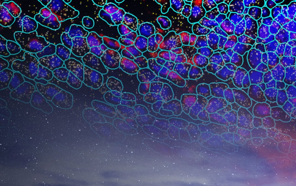
The new spatial single-cell imaging exploration instrument, the Spatial Molecular Imager, launching in 2022, complements DSP, by allowing deeper insights at both the single and subcellular levels at plex levels to ~ 1000. The spatial molecular imager is not only highly sensitive but also provides high-resolution images.
In this blog post, I’ll summarize Nanostring Senior Vice President of Research and Chief Scientific Officer, Joe Beechem’s presentation on spatial biology at the 2021 American Association for Cancer Research meeting.
GeoMx Digital Spatial Profiler to Understand Tumor Biology
GeoMx Digital Spatial Profiler (DSP) facilitates the capture of gene expression and/or protein expression alongside spatial information. You can find more information on how GeoMx DSP works and best practices for its use in studying breast cancer pathology in this Nanostring Blog article.
GeoMx DSP allows unlimited spatial multiplexing by using in-situ probes activated by light that enable ex-situ sequencing while retaining spatial information. Rather than only providing information on how cells function individually, researchers can examine how cells fit into a particular environment, and ultimately how cells function as a unit.
For example, researchers can assess gene expression in different cellular subtypes in one tumor microenvironment. GeoMx DSP can be leveraged to look at how transcriptional dynamics change in space, such as in different parts of a tumor.
Work with Dr. Jonathan Chen at the Broad Institute studying colorectal cancer revealed differences in IDO1 expression between low and high-grade core tumor regions, and also a metastatic site in the lymph nodes. Although the IDO1 pathway was low in the low-grade tumor region, it was elevated in both the high-grade and lymph nodes.
GeoMx DSP also facilitates the examination of gene expression dynamics at biologically interesting regions. For example, work by Van and Blank (2019) examined the interface between tumor and immune cells in cancer. Given the precise nature of the GeoMx DSP technology, the user can look specifically at only the interface region between the two cell types. In this example, researchers noted there was significant antigen presentation at the boundary between tumor and immune cells.
The Hubble Telescope of Spatial Biology: The Spatial Molecular Imager
Similar to the GeoMx DSP technology, samples are labeled with in-situ probes connected to a proprietary barcode. However, unlike GeoMx, the barcode is not removed but rather left in place. The precise locations of these barcodes can be read and used to infer the subcellular locations of each transcript. Transcripts can be detected, regardless of how rare the cell is in the sample – even rare transcripts in 1-cell-in-a-million can be individually quantitated.
The spatial molecular imager provides unparalleled insight into both how a cell’s individual biology shifts depending on its precise microenvironment as well as insight into the function of individual transcripts.
For example, work by Dr. Evan Newell (The Fred Hutchinson Cancer Center) examining rate T-cell populations in kidney cancer. The researchers found that cell identities and functions can change depending on where a cell is in space.
In another study, researchers showed a 3D mapping “movie” of 6 cells within a large cell number study to highlight the subcellular localization feature of SMI. This included examining over 1000 individual mRNA transcripts in space. Researchers noted that one transcript corresponding to the gene Malat1 was only found near the nucleus. This suggested that this transcript is localized to the cell nucleus.
Summary
When playing hide and seek as a child, I was limited in my ability to play the game because I was only looking in a single spatial plane. I would have been a better seeker if I thought of looking in 3D space.
Similar to my efforts as a child, looking at whole transcriptomes in their 3D context is crucial for understanding gene expression dynamics within cells. Whole transcriptome analysis in a spatial context has already been used to examine gene expression dynamics at cell-cell boundaries, and where mRNA transcripts exist within a single cell.
The Spatial Molecular Imager is launching in 2022. Once we start looking at a new spatial scale, what else will we see?
Spatial Genomics imaging is for research use only and not for use in diagnostic procedures.

