Common questions in molecular biology: How many types of cell counters are there?
Although it appears to be a simple question, there are several answers to “how many types of cell counters are there?” depending on how the word “type” is defined. Defining “type” the most broadly, there are two types, manual and automated. Manual cell counters are the original ones and still in use today. Known as hemocytometers, they require a trained technician to physically count cells under a microscope. The second type, automated, are cell counters designed to count cells quickly and autonomously. More expensive and complex than hemocytometers, automated cell counters incorporate sophisticated systems to increase reliability and throughput.
Cell counting based on method
Another answer to the question is based on the method used for counting cells. Coincidentally, the answer here is also two types: those that use microscope images for counting and those in which cells pass through a gate-type detector. Each of these groups tends to align with either the manual or automated groups. For example, image-based counters include the manual counters, plus some of the automated ones. As the name describes, image-based counters count cells in an image on a microscope. Where hemocytometers use a human scientist’s eyes for counting, automated image-based counters use imaging software connected to the microscope, allowing for quicker counts.
Image-based cell counters only count as many cells as exist in a single field of view, which can make extrapolation risky if only a small number of cells are visible.
However, whether manual or automated, image-based cell counters only count as many cells as exist in a single field of view. Accuracy can thus be affected when the machine extrapolates from the image, especially if the field of view used for counting contains a small number of cells. Some automated image counters offer solutions such as performing two counts to obtain an average, or letting users request multiple pictures from a slide. All image-based cell counters use static microscope images for counting.
Automated counters are typically in the group that tally cells as they move through a pore. Machines based on the Coulter principle contain only a counting function and operate on the premise that cells provide resistance to electrical current. Coulter counters contain two electrolyte-filled compartments separated by a pore. Cells are suspended in the electrolyte solution and flow from one compartment to the other. As each cell passes through the electrical current that crosses the pore, it provides resistance that alters the current which the machine detects and tallies.
While Coulter systems can be slower than imaging systems, they count considerably more cells (up to 20,000 animal cells) with higher accuracy from a single sample.
Cell counters available today
A more detailed answer to “how many types of cell counters are there” comes from listing the types available at the present time. The most common types are:
Image cytometers
Image cytometry is the oldest form of cell counting, using optical microscopy to statically image a large number of cells. Cells are commonly stained to enhance contrast or to detect specific molecules. Traditionally, cells are viewed in a hemocytometer to aid manual counting.
Digital cameras have made automation of image cytometers common.
Flow cytometers
Flow cytometers use the controlled flow of a solution of suspended cells to align single cells for characterization either optically or through electrical impedance detection (see Coulter counter). Specific molecules can be optically characterized by staining cells with fluorochromes similar to the ones sometimes used in image cytometry. Flow cytometers have a higher throughput than image cytometers.
Cell sorters
Cell sorters are a subset of flow cytometers that sort cells according to their innate electrical characteristics. Sorting is achieved by mechanical vibration that breaks the cell-containing fluid stream into droplets. The resulting droplets contain an electrical charge dependent on the type of cell contained within, which are counted as they are deflected by an electric field into different containers.
Time-lapse cytometers
Time-lapse cytometers facilitate continuous observation of cells inside a conventional cell culture incubator by combining time-lapse microscopy, image cytometry, and non-heat-generating light sources such as light-emitting diodes to understand cellular dynamics. Especially useful for experiments that need to avoid staining live cells that may affect the results.
Hemocytometers
Mentioned above, hemocytometers are a manual cell counting method that uses an optical microscope, a gridded microscope slide with wells of known volume, and a trained technician. Hemocytometers were the standard method of cell counting until the 1950’s.
nCounter® Pro
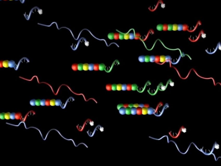
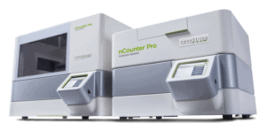
One of the more recent cell counting tools is NanoString’s nCounter® Pro Analysis System for gene expression profiling. Though not technically a cell counter per se, nCounter Pro uses a hybridization protocol of barcoded DNA probes to examine cell-specific gene expression profiles. In this way, nCounter Pro provides a relative cell abundance score that reveals the predominant cell types in a sample and which genes are playing a role in any observed effects.

Related Content


